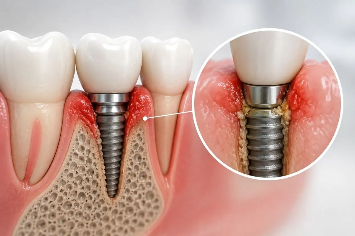
In dentistry, X-rays are an important diagnostic tool for assessing areas of the mouth that cannot be seen with the naked eye. However, not every patient needs the same imaging method. Sometimes a small dental X-ray is enough, while in other cases it may be necessary to see the jawbone, tooth roots or nerve canals in three dimensions.
This is where CBCT (Cone Beam Computed Tomography), also known as dental volumetric tomography or 3D dental tomography, comes in. CBCT makes it possible to examine the teeth and jawbones in three dimensions from different angles. However, the fact that it provides a more detailed image does not mean that it should be used at every examination or for every patient. The imaging method should be chosen according to the patient’s complaint and the planned treatment.
What Is CBCT?
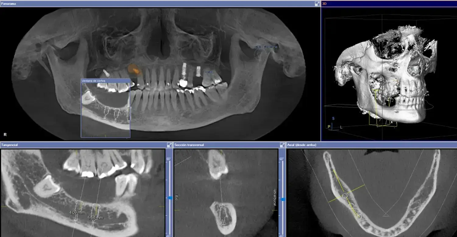
A conventional panoramic X-ray shows the teeth and jaws in a two-dimensional image. In many routine assessments, this image can provide enough information. CBCT, on the other hand, creates a three-dimensional image of the area being examined. This allows the dentist to evaluate not only left-right and up-down directions but also the depth of the structures.
For example, on a panoramic X-ray the root of a wisdom tooth and the nerve canal may appear to overlap. When necessary, three-dimensional tomography can show in more detail whether the nerve passes in front of, behind or beside the root. Similarly, in implant planning, not only the height of the bone but also its width can be assessed.
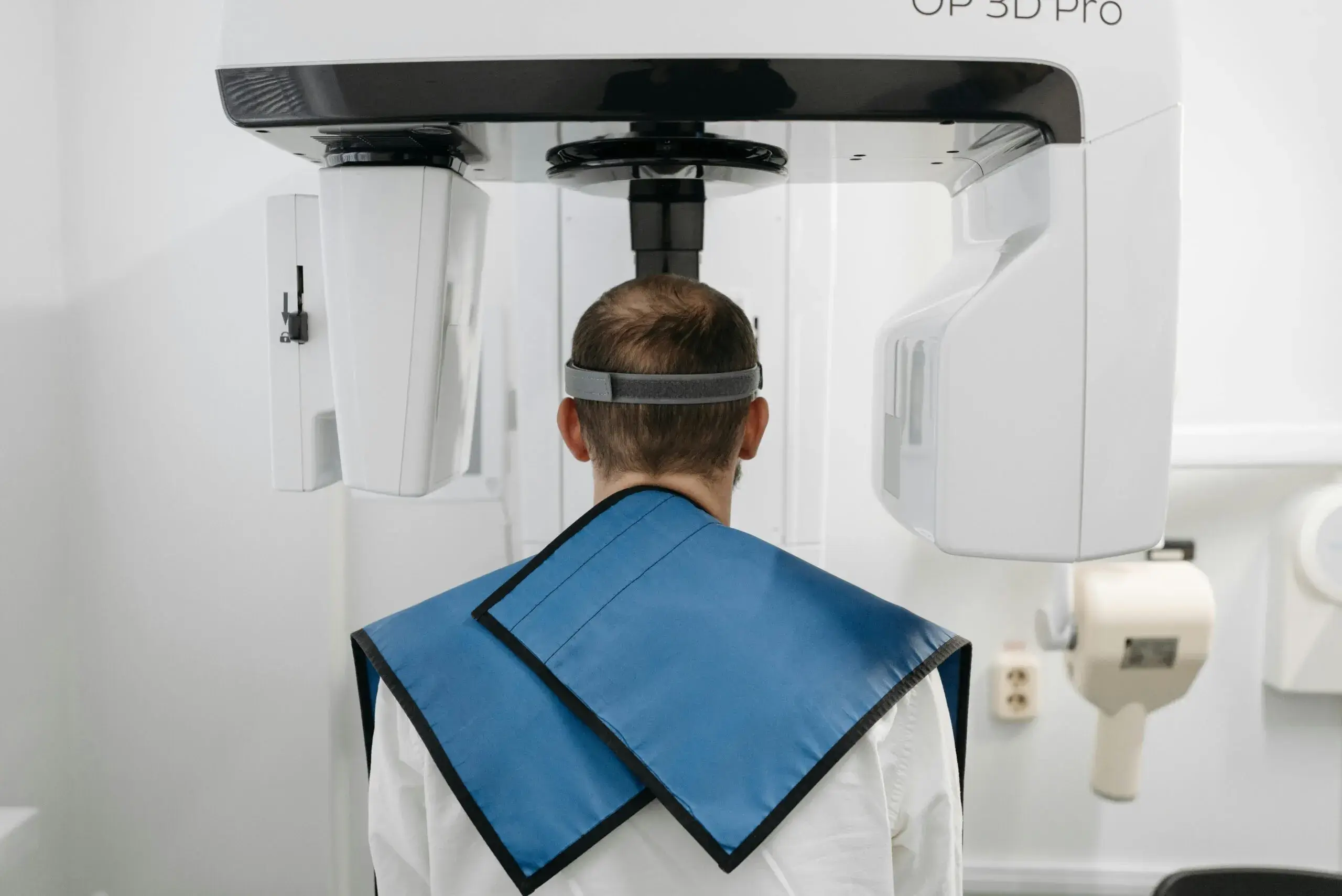
Is 3D Dental Tomography Taken for Every Patient?
No. CBCT is not an imaging method that needs to be used for everyone as part of a routine check-up. The dentist or oral surgeon first examines the patient, listens to their complaints and reviews any previous radiological images. If small dental X-rays or a panoramic X-ray provide the necessary clinical information, an additional 3D tomography scan may not be needed.
CBCT is generally preferred when a three-dimensional image could change the diagnosis or the treatment plan. This is because CBCT is an imaging method that uses X-rays. The amount of radiation used varies depending on the device, the size of the area being imaged and the scanning protocol. For this reason, the basic approach is to use the most suitable imaging method that provides the information needed, with the smallest possible imaging area.
In Which Treatments Is CBCT Used?
The treatment options for which CBCT may be preferred are as follows:
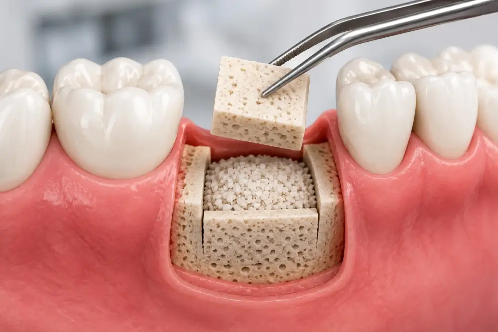
In Implant Treatment
In implant planning, the width and height of the bone and the position of neighbouring anatomical structures such as nerve canals and the sinuses are important. CBCT makes it possible to assess the three-dimensional bone structure of the area where the implant is to be placed. Especially when additional procedures such as treatment of advanced bone loss, bone augmentation or sinus lifting are being considered, three-dimensional images can provide important information for surgical planning.
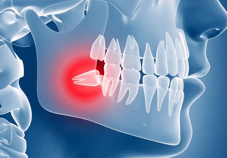
In Impacted Wisdom Teeth
In some patients, the roots of lower wisdom teeth may be very close to the nerve canal of the lower jaw. If the panoramic X-ray raises suspicion about this proximity, the three-dimensional relationship between the roots and the nerve can be examined with CBCT. This makes it easier to evaluate options such as full extraction, a controlled surgical approach or, in suitable cases, coronectomy.
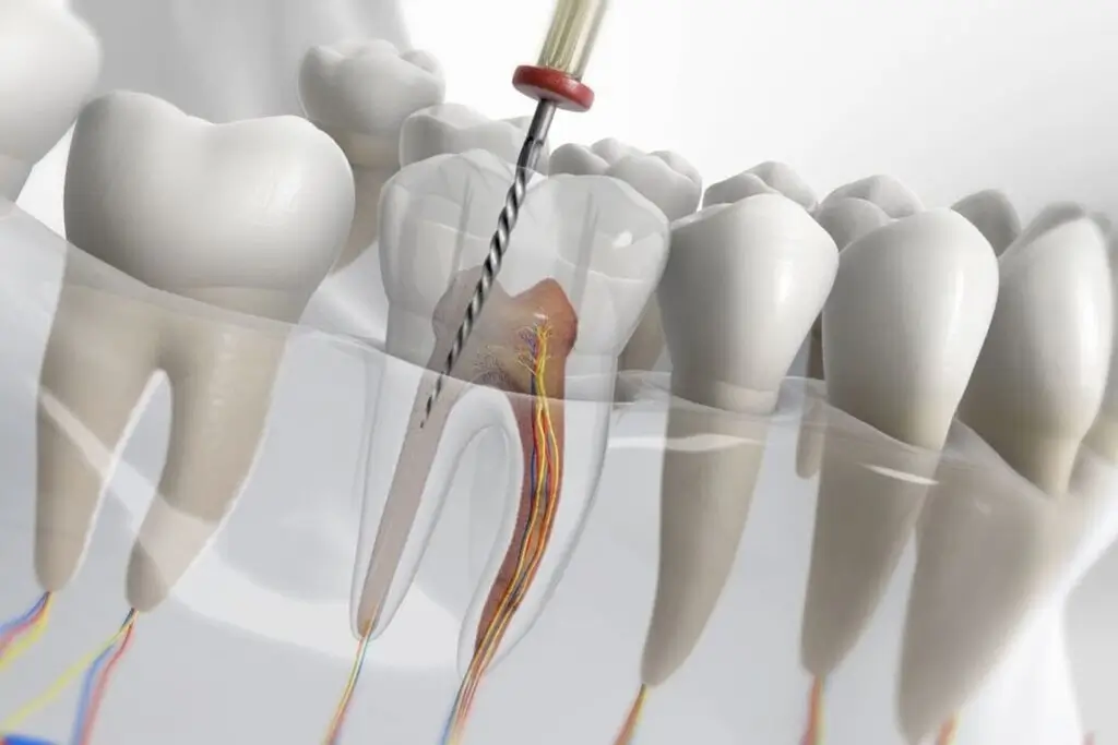
In Root Canal Treatment
Tomography is not needed for every root canal treatment. However, limited field-of-view CBCT may be used if there are complaints that cannot be explained with standard X-rays, complex root anatomy, ongoing problems in a tooth that has previously had root canal treatment, or a suspected missed canal. A three-dimensional image can help evaluate the root canals and the surrounding bone in more detail.
In Jaw Surgery and Bone Lesions
The position of cysts in the jawbone, impacted teeth and other bone-related lesions, and their relationship with the surrounding anatomical structures, can be evaluated with CBCT. There is an important distinction here: CBCT is a powerful method particularly for examining hard tissues such as teeth and bone. When soft tissues need to be evaluated in detail, other imaging methods may be required.
“More Imaging Is Not Always Better”
Dentram co-founder and oral and maxillofacial surgeon Prof. Dr. Serhat Yalçın explains the importance of using three-dimensional imaging for the right indication:
“Digital tomography gives us very valuable three-dimensional information, but requesting tomography for every patient is not the right approach. We first evaluate the clinical examination and the existing two-dimensional images. If we need more detailed information about the position of an implant, a wisdom tooth related to the nerve, advanced bone loss or a surgical lesion, we make use of CBCT. The basic principle here is to obtain the image the patient needs, in the area where it is needed. What matters is not taking more images, but answering the right clinical question with the right imaging method.”
Are CBCT and Intraoral 3D Scanning the Same Thing?
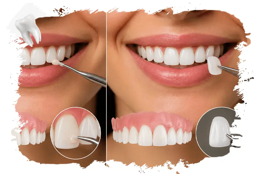
These two technologies are sometimes confused, but they are not the same. An intraoral 3D scanner optically scans the surfaces of the teeth and gums to create a digital impression and does not use ionising radiation. It can be used for procedures such as clear aligners, porcelain restorations or digital smile design.
CBCT, on the other hand, uses X-rays to show the internal structure of the teeth, roots and jawbone in three dimensions. The two technologies therefore provide different information and, when necessary, can complement each other within the same treatment plan.
How Is 3D Imaging Planned at Dentram?
At Dentram, the imaging method is chosen according to the patient’s complaint, the clinical examination findings and the planned treatment. First, it is assessed whether the existing radiographs provide enough information. When a three-dimensional examination is needed, dental volumetric tomography is used to examine the bone structure and anatomical relationships of the relevant area in detail.
In different areas, from implant surgery to impacted teeth and from advanced bone augmentation to complex root canal treatments, CBCT can make diagnosis and treatment planning more controlled and predictable when used for the right patient. What matters is not applying the most advanced imaging method to every patient, but obtaining the information needed with the most suitable method.