In some patients, the roots of impacted wisdom teeth in the lower jaw can sit very close to the nerve canal that carries sensation to the lower lip and chin. If the tooth roots and the nerve canal appear to overlap on a panoramic dental X-ray, patients usually have the same question in mind: “Will the nerve be damaged if this tooth is removed?”
A tooth being close to the nerve does not necessarily mean that nerve damage will occur or that the tooth cannot be removed. However, it does show that the procedure should not be planned like a standard extraction. In such cases, the root structure of the tooth, the position of the nerve canal and the surrounding bone are examined in detail. Where necessary, 3D dental tomography is used to create a surgical roadmap specific to the patient.
What Does It Mean If a Wisdom Tooth Is Close to the Nerve?
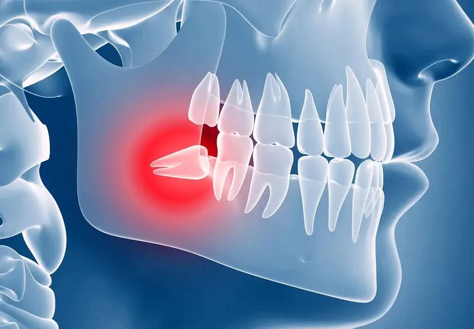
Inside the lower jaw there is an anatomical structure called the mandibular canal. The inferior alveolar nerve, which runs through this canal, provides sensation to the lower lip, chin, lower teeth and surrounding gum tissue. In some impacted wisdom teeth, the roots may develop directly above, beside or around this canal. When there is not enough bone between the roots and the nerve, the risk of the nerve being stretched, compressed or injured during extraction may increase.
If the nerve is affected, numbness, tingling or an altered sensation may occur in the lower lip or chin. A significant proportion of these changes may be temporary. However, because there is also a risk of permanent changes in sensation in cases closely related to the nerve, assessment before surgery is very important. The nerve in question is not one that moves the facial muscles. Any possible effect is mostly related to sensation in the lower lip and chin.
Is a Panoramic X-ray Enough?
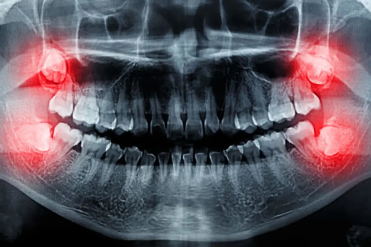
Wisdom teeth are usually first assessed with a panoramic dental X-ray. This X-ray shows the direction in which the tooth is impacted, the general shape of the roots, its relationship with neighbouring teeth and how close it is to the nerve canal. However, a panoramic image is two-dimensional. The tooth root and the nerve canal may appear to overlap on the X-ray, but it is not always clear whether the canal passes beside the root, on its inner side or on its outer side.
If the panoramic X-ray shows signs suggesting a close relationship with the nerve, or if the image does not provide enough information for surgical planning, 3D dental tomography may be requested. Tomography is not needed for every wisdom tooth extraction. The imaging decision is made based on the patient’s existing X-ray and the clinical examination findings.
What Does 3D Dental Tomography Show?
3D dental tomography, also known as cone beam computed tomography, allows the teeth and jaw structures to be examined in three dimensions. This way, the surgeon can evaluate the relationship between the tooth roots and the nerve canal from different angles. Tomography makes it possible to answer the following questions more clearly:
- On which side of the tooth root does the nerve canal pass?
- Is there a layer of bone between the roots and the canal?
- Are the roots curved, separated or shaped around the nerve?
- From which direction is it safer to approach the tooth for removal?
- Should a different surgical method be considered instead of a full extraction?
3D imaging does not eliminate the risk completely. However, by showing the anatomical relationship more clearly, it helps the surgeon plan in advance which method to use, how the tooth will be divided into sections and how much bone will be removed.
How Is a Wisdom Tooth Close to the Nerve Removed?
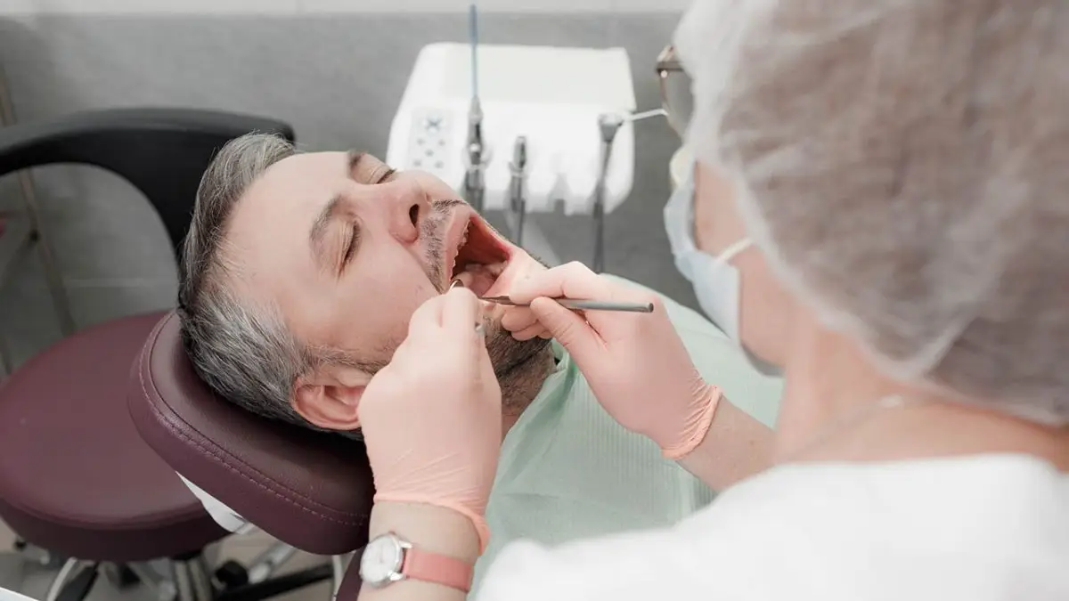
The extraction method is not the same for every patient. Depending on the position of the tooth, the gum can be opened in a controlled way and a limited amount of bone over the tooth can be removed. Instead of forcing the tooth out in one piece, the crown and root sections can be divided into smaller pieces. This approach can reduce the force needed to remove the tooth. The aim is to avoid sudden and uncontrolled movements, especially with roots close to the nerve canal.
Based on the tomography images, the surgeon decides from which direction the tooth will be removed and which root section will be addressed. The procedure can be performed under local anaesthesia. Depending on the patient’s level of anxiety, the extent of the operation and their general health, other anaesthesia or sedation options may also be considered.
What Is Coronectomy and When Is It Performed?
In some patients, the roots of the wisdom tooth may be in very close contact with the nerve canal. In suitable cases where removing the whole tooth carries a high risk for the nerve, a method called coronectomy may be considered. In coronectomy, the upper part of the tooth that causes problems in the mouth, in other words the crown, is removed, while the roots close to the nerve are deliberately left in the jawbone. The aim is to reduce the risk of injury that could occur while trying to separate the roots from the nerve.
This method is not suitable for every patient. If there is an infection around the tooth or roots, or if there is a condition that prevents the roots from being safely left in place, a full extraction of the tooth may be necessary. Because roots left in the jaw may move later, it is important for patients who have had a coronectomy to be followed up regularly. In rare cases, the roots may need to be treated again at a later stage.
Does Being Close to the Nerve Prevent Extraction?
Dentram co-founder and oral and maxillofacial surgeon Prof. Dr. Serhat Yalçın explains the importance of detailed imaging for wisdom teeth close to the nerve:
“A wisdom tooth appearing close to the nerve canal on a panoramic X-ray does not mean that the tooth cannot be removed. What matters here is correctly assessing the three-dimensional relationship between the roots and the nerve. Where necessary, we use dental tomography to examine on which side of the tooth the nerve passes, whether there is protective bone tissue in between and how the surgical approach should be planned. In some cases we can carry out a full extraction by dividing the tooth into sections in a controlled way, while in suitable patients with a high risk of nerve damage we can consider coronectomy. Surgical success depends less on removing the tooth quickly than on personalised planning that respects the anatomical structures.”
What Happens If Nerve Damage Occurs?
After the extraction of wisdom teeth close to the nerve, numbness, tingling or a change in sensation may rarely be felt in the lower lip or chin. This does not always mean that there is permanent nerve damage. Depending on how much the nerve has been affected, the loss of sensation may decrease over time, and in many patients sensation gradually returns.
The recovery time is not the same for every patient. In some patients the changes improve quickly, while in others recovery may take longer. Even in cases that do not fully recover, the feeling of numbness may become less noticeable over time with the contribution of the surrounding nerves. In some patients this process can take up to 1–2 years.
When the nerve has been affected, B complex vitamins or photobiomodulation, also known as low-level laser therapy, may be used to support the healing process, depending on the dentist’s assessment. Photobiomodulation is a non-surgical method that aims to support the healing of nerve tissue through the application of low-energy light.
If numbness and changes in sensation persist for a long time, or if a serious nerve injury is suspected, a more detailed assessment is carried out. In selected cases where it is considered necessary, microsurgical treatment options may also be considered.
How Does the Assessment Process Work at Dentram?
At Dentram, impacted wisdom teeth close to the nerve are first assessed with a clinical examination and a panoramic X-ray. If the relationship between the tooth roots and the nerve canal is not clear, or if close contact is suspected, 3D dental tomography is used.
The angle of impaction, the root structure, the position of the nerve canal and the surrounding bone are examined together on the images. Whether a full extraction, controlled sectioning or coronectomy is the most suitable option is decided individually. The possible risks of the procedure, the alternatives and the follow-up process are explained clearly to the patient.
If you have been told that your wisdom tooth is close to the nerve, this does not mean that treatment is not possible. With the right imaging, an experienced surgical assessment and personalised planning, the most suitable method of intervention can be determined. You can book an assessment appointment with Dentram’s oral and maxillofacial surgeons to get detailed information about your tooth’s relationship with the nerve and your treatment options.