
What Are NICO, Cavitation, and FDOJ? How Are They Treated and Prevented?
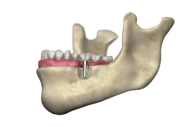
One of the most important yet least known topics in biological dentistry is the hidden lesions that occur in the jawbone, referred to as NICO, FDOJ, or “cavitations.”
Cavitations are a subject rarely emphasized in conventional dental education. However, current scientific studies show that these lesions may be associated with many serious health conditions, ranging from unexplained trigeminal neuralgia (severe facial pain) to systemic and autoimmune disorders. Especially if you have had a difficult wisdom tooth extraction in the past or are planning an oral surgery procedure in the near future, it is of great importance to be informed about these “silent inflammation” areas in the jawbone.
What Do NICO, FDOJ, and Cavitation Mean?
Cavitation (cavity) is actually a well-known term in orthopedics, used to describe lesions found in knee and hip bones. Although these cavities in the jawbone were first observed as early as the 1920s, the first person to study the subject in detail was oral pathology specialist Jerry Bouquot. Bouquot identified these jawbone lesions particularly in patients suffering from unexplained nerve-related facial pain (neuralgia) and named the condition NICO (Neuralgia-Inducing Cavitational Osteonecrosis). Here, “osteonecrosis” refers to dead bone tissue, and “cavitation” refers to the hollow space within the bone.
In the following years, Johann Lechner, one of the pioneering figures in biological dentistry, discovered that these lesions were not exclusive to neuralgia patients but could silently persist in many individuals without causing any pain symptoms. When Lechner surgically opened the cavitation areas, instead of healthy bone tissue, he encountered degenerative tissue that easily crumbled, was soft, and contained fat droplets. For this reason, he updated the name of the condition to FDOJ (Fatty Degenerative Osteolysis of the Jawbone). According to Bouquot, the cause of this dense fat cell accumulation is insufficient blood supply (circulation) in the area.
Why Are NICO, FDOJ, and Cavitations So Important?
Some of these lesions cause severe pain in patients, leading to unnecessary root canal treatments, extraction of healthy teeth, or the use of heavy neurological medications. However, the real danger lies hidden in “silent” cavitations that show no symptoms.
Lechner’s tissue analyses revealed a striking finding: Inside the cavitations, cytokines such as TNF-α and IL-6, which we would expect to see in a typical inflammation, were virtually absent. Instead, an inflammatory cytokine (chemokine) called RANTES (CCL5) was found, reaching levels up to 35 times above normal. This condition is called “silent inflammation.” The absence of conventional inflammatory markers prevents the immune system from detecting the problem, allowing it to remain hidden for years.
What Is the RANTES / CCL5 Cytokine?
RANTES is a signaling molecule that recruits immune cells to inflamed areas. While beneficial in acute situations, it appears as a factor that increases cellular damage and facilitates disease progression (metastasis, etc.) in conditions such as rheumatoid arthritis, multiple sclerosis (MS), chronic fatigue syndrome, and even breast, stomach, and pancreatic cancers. A NICO lesion in the jawbone acts as a source that continuously pumps RANTES into the body, fueling systemic inflammation. Indeed, when cavitations are surgically cleaned, a significant drop in RANTES levels has been observed in patients’ blood.
How Is It Diagnosed?
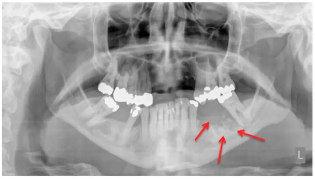
NICO and FDOJ lesions are usually missed on conventional two-dimensional X-rays. While signs may be visible in advanced cases, 3D Dental Cone Beam Computed Tomography (CBCT) is essential for definitive diagnosis. Additionally, specialized dental ultrasound devices (CaviTAU, etc.) that measure bone density are now being used (particularly becoming widespread in Europe) to detect these fatty degeneration areas.
How Are Cavitations Treated?
Cavitation areas are avascular (lacking blood supply) regions. Since there is no circulation, it is physiologically impossible for any antibiotic or medication taken orally to reach this area and promote healing.
The only rational treatment method is surgical curettage. The affected bone area is surgically opened, the necrotic fatty tissue inside is physically scraped (curetted) and completely removed, and fresh bleeding is induced in the area. Within the framework of Dentram biological dentistry protocols, medical ozone is applied to eliminate pathogens in the cleaned area, and PRF (Platelet Rich Fibrin) membranes obtained from the patient’s own blood are placed in the area to accelerate healing, completing the operation.
What Causes NICO and FDOJ?
95% of cavitations occur following tooth extractions (especially impacted wisdom teeth). The main factors that disrupt the healing process and cause fat tissue to form instead of bone are as follows:
- Coagulation Disorders: Hypercoagulability (excessive clotting tendency) in the patient’s blood disrupts microcirculation in the extraction socket, preventing adequate bone nourishment.
- Stem Cell Differentiation (Vitamin D and K2 Deficiency): After extraction, stem cells in the area require certain signals to differentiate into bone tissue. Vitamin D deficiency, osteocalcin insufficiency (which requires Vitamin K2), diabetes, and hormonal imbalances (estrogen deficiency, etc.) cause stem cells to differentiate into fat cells (adipocytes) instead of bone.
- VDR (Vitamin D Receptor) Blockage: Hidden infections such as Epstein-Barr, Lyme (Borreliosis), Chlamydia, and Cytomegalovirus can inactivate the Vitamin D receptors (VDR) in cells. In this case, even though the Vitamin D level measured in the blood appears adequate, it cannot enter the cells and healthy bone metabolism cannot take place.
- Additionally, autoimmune diseases, hypothyroidism, and heavy metal toxicity are also among the factors that impair healing.
How Can NICO and Cavitations Be Prevented?
The most effective way to prevent these lesions is to strictly follow biological healing protocols before, during, and after tooth extraction.
1. Pre-Extraction Preparation (Systemic Support)
- If there is no urgent infection, the extraction procedure should be scheduled during the period when the patient’s overall health is most stable.
- Starting supplementation with Vitamin D3-K2, Magnesium, Zinc, and high-dose Vitamin C weeks before the operation is essential for bone healing.
- An anti-inflammatory diet, free from packaged and processed foods, should be adopted; quality protein (minimum 0.8g per kilogram of body weight) should be consumed to support tissue repair.
- Regular exercise should be performed to improve microcirculation, and attention should be paid to sleep patterns (circadian rhythm) for hormonal balance.
2. During Tooth Extraction (Surgical Protocol)
- After the tooth is extracted, the thin membrane (periodontal ligament) that connects the tooth to the bone must be completely removed. We perform this procedure using piezosurgery devices that operate with ultrasonic vibrations and do not damage surrounding tissues.
- Local anesthetics containing vasoconstrictors (adrenaline, which constricts blood vessels) can increase the risk of cavitation by reducing blood supply to the area; therefore, the choice of anesthetic must be made carefully according to the patient’s condition.
- The extraction socket should be disinfected with Ozone gas, and PRF (a stem cell membrane containing growth factors) prepared from the patient’s own blood must be placed in the extraction socket.
3. Post-Extraction Care
- The first and most important step of wound healing is the preservation of the blood clot that forms in the extraction socket. Rinsing the mouth and spitting must absolutely be avoided after the procedure.
- Using straws, which create a vacuum effect that can dislodge the clot, and smoking are strictly prohibited.
- After the first 24 hours, very gentle mouth baths with salt water or Lugol’s solution can be performed to support cell regeneration.
- Physical activity should be avoided for the first 3-4 days, and rest should be prioritized. To increase tissue oxygenation, intravenous (IV) high-dose Vitamin C support should be utilized (especially upon physician recommendation).